LooksLab guide
Flat maxilla: How to Check and What Can Change
Does your midface look flat? Learn what a flat maxilla means, how to check your profile, and what orthodontics, surgery and filler can actually change.
A flat maxilla is an informal description of an upper jaw that looks short on forward projection, although a flat-looking midface does not establish a jaw diagnosis. In adults, mewing cannot reshape it; surgery can reposition the upper jaw, while filler changes soft-tissue contour. Jaw terminology, mewing evidence, jaw surgery, filler guidance.
This guide is for adults 18+. Start by naming the area that bothers you: beside the nose, the outer cheeks, beneath the eyes or the overall profile. Those are different questions, and you do not have to decide on a treatment to ask them.

Fictional illustration, not a patient or a treatment result.
What does a flat maxilla actually mean?
Your maxilla is your upper jaw. It holds the upper teeth and contributes to the mouth, nasal cavity and eye sockets. It connects with the zygomatic bone, or cheekbone, but they are distinct bones. Maxilla anatomy.
Two clinical terms help untangle the search results:
- Maxillary hypoplasia: underdevelopment or reduced size of the upper jaw.
- Maxillary retrusion: a backward position relative to the surrounding facial skeleton.
Size and position are related, but they are not interchangeable. Neither term means that every part of the midface sits equally far back. Clinical definitions.
“Forward maxilla” describes the opposite direction people are asking about. It is not a target you need to maximize. A useful consultation question is whether your upper jaw, lower jaw and teeth fit together, and which region accounts for the appearance you notice.
Do I have a flat maxilla, or just flat cheekbones?
Separate the location from the label. The maxilla supports the central midface; the zygoma supports its outer portion. The nose base and upper lip also involve skeletal support and covering tissues. Regional anatomy, nasal-base and lip support.
Use this table to choose a question, not to diagnose yourself. More than one row may describe your concern.
| What you notice | What needs separating | Useful question |
|---|---|---|
| “It looks flat beside my nose; my upper lip sits back” | Upper-jaw position, the tooth-bearing region and lip thickness | Is this mainly skeletal position, dental support or soft-tissue contour? |
| “My cheekbones look flat or low” | The outer cheekbone from the central upper-jaw region | Which part of my cheek is the concern, and would a jaw movement even address it? |
| “My under-eyes look hollow” | Under-eye contour from a diagnosis of upper-jaw deficiency | Is the concern a hollow or the support beneath the eye? |
| “My chin looks weak and my whole profile looks off” | The relationship between the upper and lower jaws | Which jaw, if either, is out of position? |
| “My midface looks flatter than it used to” | Changes in soft tissues and skeletal support with age | What has changed, and what older photos or history would help assess it? |
The jaw-relationship distinction comes from the clinical examination chapter; the aging distinction comes from a review of facial skeletal changes.
For an eye-area concern, our guide to recessed infraorbitals and under-eye support covers that region in detail. Keep it separate from the question of where your upper teeth and jaw sit.
What if my bite seems normal?
Record that too. “My cheeks look flat” describes appearance; “my teeth meet comfortably” describes your experience of your bite. Neither observation alone identifies upper-jaw position. The clinical distinction between the whole midface and the maxilla is why an outer-cheek concern should not automatically become a jaw-surgery question. Ask for the region to be identified before discussing a procedure.
How can I check my side profile without guessing?
Make one repeatable photo record and write down what you see. The goal is a useful question for an appointment, not a pass/fail picture.
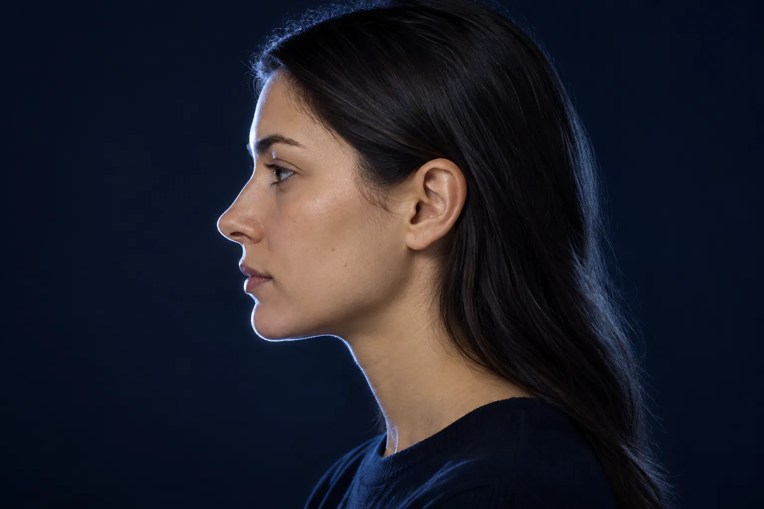
Fictional illustration of a level profile. For your own photo, point an eye-level camera at the side of your face.
- Set a steady camera at eye level. Use a timer or helper. Avoid an extreme close-up; leave room around your head.
- Take a true side view. Face straight ahead while the camera looks at the side of your face. Keep your nose, lips and chin visible, your head level and your expression relaxed. Do not jut your jaw or lift your chin.
- Take a frontal photo in even light. Turn off beauty filters. If lighting is part of your question, take an optional second frontal photo under ordinary overhead light, keeping everything else the same.
- Save the setup. Note the camera, distance, height, light and expression so you can repeat it.
The guide to taking consistent front and side photos explains the capture setup. This worksheet is an observation aid, not a clinical test:
| Record | Your note |
|---|---|
| Region noticed | Beside nose, upper lip, outer cheek, under-eye or whole profile |
| Change with light or angle | Stronger, weaker or similar; name the setup |
| Bite or chewing concerns | What feels different, if anything, and when you noticed it |
| Question for clinician | What would you like clarified? |
A hypothetical entry: “The area beside my nose looks flat in profile. I have not noticed a bite change. Is this mainly jaw position or surface contour?” That is enough to start a conversation. You do not need to arrive with a diagnosis or a proposed operation.
What can the photo actually tell me?
It records visible contour. It cannot measure the hidden skeletal landmarks used in a cephalometric assessment. A clinical case report illustrates the difference: assessment included a bite examination, lateral cephalograms and multiple skeletal and dental measurements.
An orthodontist or oral and maxillofacial surgeon interprets that examination and, when indicated, a cephalometric X-ray to assess jaw position. Do not estimate an X-ray angle from a selfie, use a forehead line as a diagnostic cutoff, or press beneath your eyelid to look for bone.
Bring persistent bite or chewing concerns to an orthodontist. Ask what the findings mean together and whether the issue is dental, skeletal or mainly contour.
Why does my midface look flat?
There is no single cause you can read from the word “flat.” The anatomy above separates several possible contributors; your history matters too.
Developmental deficiency is one clinical possibility. Maxillary hypoplasia is described in people with cleft lip and palate, for example. That is a recognized clinical context, not a reason to assign a developmental disorder to someone with flat-looking cheeks. LeFort I review.
Aging is a different context. A narrative review describes selective bone resorption and loss of maxillary projection with age, contributing to changes around the nose and under-eyes. This does not establish why a young adult has a particular contour, or show that exercises can grow the jaw forward. Mendelson and Wong.
Childhood mouth breathing is not a personal diagnosis. A review found inconsistent findings and methodological problems in research on breathing and facial development. It cannot tell you that a childhood habit caused your face shape or that changing your breathing now will reverse it. Lin et al..
Can you fix a flat maxilla without surgery?
It depends on whether “fix” means changing tooth position, adding contour or moving the jaw itself. Those outcomes need separate discussions.
For growing children, clinicians may consider reverse-pull facemask treatment while the surrounding sutures are still developing. The pediatric case report's discussion explains that growth-dependent timing. It does not establish adult results or a universal age cutoff. A child's clinical appliance is not evidence for an adult home routine.
For adults, braces move teeth; that is different from surgically repositioning the tooth-bearing upper jaw. The LeFort I review emphasizes coordinating skeletal movements with dental treatment. If braces or aligners are proposed, ask what would change in the teeth, what would change in the jaw, and what risks or compromises the plan involves.
For surface contour, filler is a nonsurgical option; cheek implants and fat grafting are surgical options. They add volume rather than reposition the tooth-bearing jaw. FDA, ASPS.
Does mewing fix a recessed maxilla?
No. Mewing does not work to reshape an adult maxilla. Cleveland Clinic explains that tongue exercises lack the strength and specificity to significantly change facial or dental features. Cleveland Clinic's evidence explanation.
Do not use chewing gum, jaw exercises, face pulling or bone smashing as a maxilla treatment. A change in how a posed photo looks is not evidence of skeletal advancement. Use the repeatable record above rather than trying to train toward a different profile.
What do surgery, filler and implants actually change?
These are treatment categories to understand before a consultation, not recommendations to get a procedure. The useful comparison is the tissue being changed and the risks involved.
| Option | What it changes | Main limits and risks |
|---|---|---|
| LeFort I jaw surgery | Repositions the tooth-bearing upper jaw; advancement is one possible movement | Does not automatically advance the entire cheekbone and orbital-rim complex. Jaw-surgery risks include bleeding, infection, dental damage and persistent numbness. |
| Dermal filler | Adds soft-tissue volume in selected areas | Does not reposition the jaw or correct its bite relationship. Rare vascular complications can cause tissue death, blindness or stroke. |
| Cheek implants | Surgically add cheek volume | Augmentation is different from jaw repositioning. Risks include implant displacement, infection, altered sensation and possible revision. |
| Fat grafting | Surgically transfers fat to add cheek volume | Does not move the tooth-bearing jaw. Risks include infection, altered sensation and, rarely, blindness. |
Sources: LeFort I target, jaw-surgery risks, filler benefits and risks, cheek augmentation and augmentation risks.
How much recovery does jaw surgery involve?
Cleveland Clinic gives these general jaw-surgery milestones:
- Return to a daily routine: often three to four weeks.
- Initial healing: about six weeks.
- Complete jaw healing: may take a year.
These are general guidance, not a recovery forecast for an isolated LeFort I operation. Your operation and individual recovery matter. Returning to daily life and complete healing are different milestones. Cleveland Clinic recovery guidance.
For filler, duration depends on the material and injection area; absorbable products lose their effect over time. FDA approvals are specific to the use and product, with the cited approved uses for adults 22 or older. This article's 18+ audience does not make every reader eligible. FDA guidance.
Before considering structural treatment, see an orthodontist or oral and maxillofacial surgeon. Ask which region would change, how your bite would be managed, and what recovery and follow-up the specific plan requires. For contour procedures, ask the treating clinician about the exact product or operation, expected duration and complications.
Can a face-analysis tool measure my maxilla?
LooksLab's free browser tools analyze a front photo, not a side profile. None measures forward maxilla projection, bite or cheekbone depth.
The golden ratio face calculator compares frontal proportions around the eyes, nose, mouth and chin. Its eye–nose–mouth ratio describes relationships in the front view; it does not measure how far the upper jaw projects forward.
The free front-photo attractiveness test shows a formula and factor breakdown using visible proportions, feature placement and symmetry. Its midface proportion is a frontal vertical ratio, not a jaw-depth measurement. Skin and facial fat are unmeasured in this web test. Scores are app estimates from one photo.
Use these tools if your question is about frontal proportions. Keep the profile worksheet for your side-view observations and a clinical assessment for the bone-and-bite question.
What can I work on now?
Choose one next step that matches your concern:
- For an unclear photo: make the consistent record above and write one question.
- For bite or chewing concerns: arrange an orthodontic assessment and bring your notes.
- For everyday appearance: choose a styling or skin-care priority from our guide to what a routine can actually change.
You can leave the procedure question open while you work on an appearance goal you have chosen. There is no need to make your entire routine about one profile feature.
To organize those goals, start your LooksLab plan: it gives you AI face analysis and a week-by-week glow-up plan built around what can change.